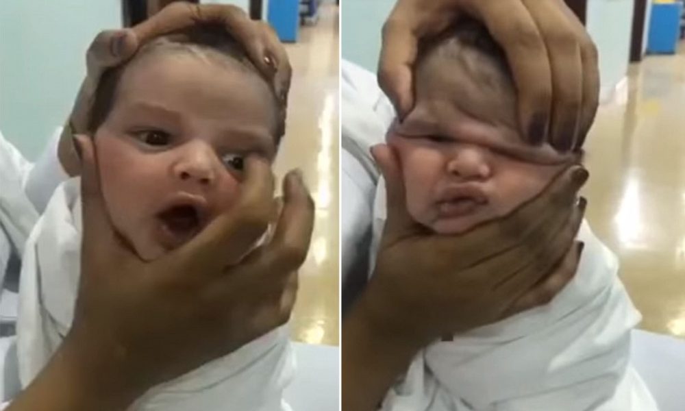
A baby girl has been born with two mouths due to a disease that’s only been seen in 35 known occurrences since the year 1900. The condition is extremely rare and has only been observed 35 times in over 100 years…Click Here To Continue Reading>> …Click Here To Continue Reading>>
When the anomaly was discovered during a scan performed during the mother’s third trimester, doctors were perplexed. At first, they considered a variety of diagnoses, including a cyst, an underlying bone problem, and teratoma, which occurs when a twin absorbs another during development in the womb.
The baby was born in Charleston, South Carolina, and physicians discovered what they characterized as a duplicated oral cavity or second mouth when she was examined. This contained an additional lip, a set of six teeth, and even a little tongue that moved in sync with the tongue in her main mouth at the moment of birth.
Despite the fact that the condition appeared to be safe, they decided to undertake an operation to remove the additional function. The irregularity was discovered by doctors during week 28 of pregnancy, and they initially suspected it to be a cyst or a tumor. Medical professionals in Charleston, South Carolina, determined that the 0.8-inch growth was actually an additional mouth when the tiny girl was delivered there.
According to the doctors who published their findings in the journal “BMJ Case Reports,” her second mouth was not connected to her main mouth, and she was unable to breathe, eat, and drink regularly. They did, however, point out that it would occasionally exude a clear liquid, possibly saliva, and that a rough surface would emerge around it at other times.
The small child was admitted to the hospital for serious surgery in order to have the additional organ taken out. During this procedure, she had to have her lower jaw, known as the mandible, drilled down to remove excess bone that was supporting her teeth for the second mouth.
The doctors noted that the child had developed a minor puffiness of the right face near the surgical incision following the operation, which they said was normal. A scan was performed, and the results revealed a fluid collection. According to the doctor’s study, the fullness resolved over a period of several months, and she did not require any more treatment.
They explained that after six months, the wounds were completely healed, and the patient was able to feed without any trouble. However, the physicians noted that she was unable to pull her right lower lip downwards, which could indicate that the muscles in that area had stopped working altogether.
Diprosopus, which means “two faces” in Greek, is an extremely rare illness that’s been observed in chickens, sheep, cats, and other animals, among other things. Scientists believe it was caused by the sonic the hedgehog (SHH) gene, which interferes with the construction of the skull during embryonic development. The rare “two faces” disorder, diprosopus, is one of the rarest of the rare.
Craniofacial duplication, which is Greek for “two faces,” is an extremely rare genetic condition. It’s a congenital abnormality that results in the duplication of some facial characteristics. It’s expected that a newborn born with this syndrome would have one body and normal limbs, but his or her facial characteristics will be replicated to various degrees. In milder cases, a newborn may be born with two noses and four eyes that are spread widely apart from one another.
However, in extreme circumstances, the complete face of a baby could be replicated. In many instances, newborns are born without a brain and with severe heart problems. The vast majority of newborns diagnosed with diprosopus are stillborn, and there have only been a few hundred cases reported worldwide.
The BMJ Case Reports journal has released a comprehensive medical report on the girl’s situation. Specifically, it reads as follows: ‘During prenatal imaging, a right mandibular tumor was discovered in a baby girl who was transferred to a medical clinic for evaluation. There was a one to two-centimeter lump along the right jaw, and there appeared to be remnants of a vestige of an oral cavity.
On physical examination, teeth-like tissue that resembled the vermilion and a rudimentary tongue looked to be innervated and moved in sync with the mouth movement. Examination of the jaw after birth revealed a soft tissue mass of the right mandibular body that was partially osseous and contained uninterrupted teeth, as confirmed by MRI and CT scans. Six months after birth, she was sent to the operating room for a large-scale excision and reconstruction. READ FULL STORY HERE>>>CLICK HERE TO CONTINUE READING>>>
She recovered quickly after surgery and was able to feed herself without problems. Skeletal duplication, which includes duplication of stomatodil structures or the presence of diprosopus, is a rare disorder that manifests itself in a variety of ways. Associated syndromes should be excluded in the case of suspected craniofacial duplication, and adequate imaging should be performed to identify the extent to which surrounding tissues have been affected.
This information will ultimately be used to guide surgical strategy. Approximately 35 occurrences of diprosopus (duplication of craniofacial structures) have been described in the literature since 1900, making it one of the most unusual conditions known. It’s possible to have craniofacial duplication in combination with other congenital defects, resulting in a wide range of symptoms, ranging from complete facial duplication to partial duplication of facial components.
Typically, the maxilla, mandible, and oral cavity are the most affected areas when partial duplication takes place. Additionally, cerebral involvement is possible, with the pituitary gland duplicating being the least severe kind. Females are more likely than males to be affected by the illness, but the mechanisms that influence this demographic are still being investigated. Cleft lip and palate, cleft palate syndrome, and the Pierre Robin sequence are all common comorbidities linked with craniofacial duplication.
During the third trimester of pregnancy, a right mandibular mass was seen on prenatal ultrasonography, prompting the referral to another medical center. The original differential diagnosis included a variety of conditions, such as congenital cyst, sinus teratoma, fibrous dysplasia, and foregut duplication, among others. There was no evidence of in-utero teratogen exposure, and there was no family history of face deformities.
A Caucasian girl was born to a healthy mother at 40 weeks and four days after the birth of her daughter through an uneventful spontaneous vaginal delivery. There were no indicators of respiratory distress, and there was no reason to be concerned about the bulk getting into the airways. On inspection of the infant, it was discovered that the right body of the mandible had a one to two-centimeter fullness, with the tongue being displaced to the left over the course of the examination.
In the right oral commissure, there was a tiny sinus tract with vermilion-appearing mucosa surrounding that. It was one centimeter inferior and lateral to the commissure. The sinus tract measured around 13 millimeters in depth and was located next to the mandibular mass, with no obvious contact in the mouth cavity or other structures. The mandibular level ridge had been expanded several centimeters on the left side, and she’d lost the capacity to compress the lower lip on the right side of the face.
Other than that, the rest of the head and neck cheek was uneventful. After admission to the newborn nursery, the patient showed no signs of respiratory distress and was able to consume enough food before being transferred to the general ward for discharge after two weeks of age.
The infant appeared healthy and was feeding and gaining weight normally, with no signs of oral incompetence, according to the doctor who examined her at the time. It was discovered that the exterior component of the mass periodically developed a raw surface at the skin level that drained a clear serous fluid that appeared to be saliva-like in appearance.
The fluid, on the other hand, was not subjected to any tests. When the infant was feeding, a little accessory tongue seemed to protrude from the aperture of the sinus canal and was observed to move in synchronization with the oral tongue. When the patient was six months old, she was sent to the operating room for excision of the duplicated mandible, bone contouring of the jaw, and repair of the soft tissue defect using nearby tissue transfer, among other procedures.
A plane between the uninvolved soft tissue and the duplicate oral cavity was created with the use of a combination of blunt and sharp dissection techniques. The mucosal lining and minor salivary glands connected with it were removed in one piece and traced all the way to the jaw. The mucosal lining extended onto the mandible and resembled the mucosa overlaying the level arch in its appearance and texture.
When this was pulled away from the mandible, the underlying bone showed six primary teeth that were oriented toward the duplicate oral cavity of the original patient. It was decided to extract the accessory teeth, and the sockets were drilled down to shape the jaw and remove any remaining dental tissue. It was important to avoid removing any tooth buds that were deemed to be a part of her natural mandible. The facial nerve was preserved by the use of intraoperative nerve monitoring.
In order to close the remaining soft tissue defect, an advancement flap was used, which had an area of approximately 83.3 centimeters squared. Following the surgery, pathology revealed benign squamous mucosa, salivary gland, cortical bone, skeletal muscle, and dental pulp with six benign molar teeth, which was removed during the operation.


 HEALTH & LIFESTYLE11 months ago
 IN-THE-NEWS6 months ago
 SPORTS9 months ago
 SPORTS10 months ago
 SPORTS11 months ago
 IN-THE-NEWS6 months ago
 SPORTS9 months ago
 METRO10 months ago


